Introduction
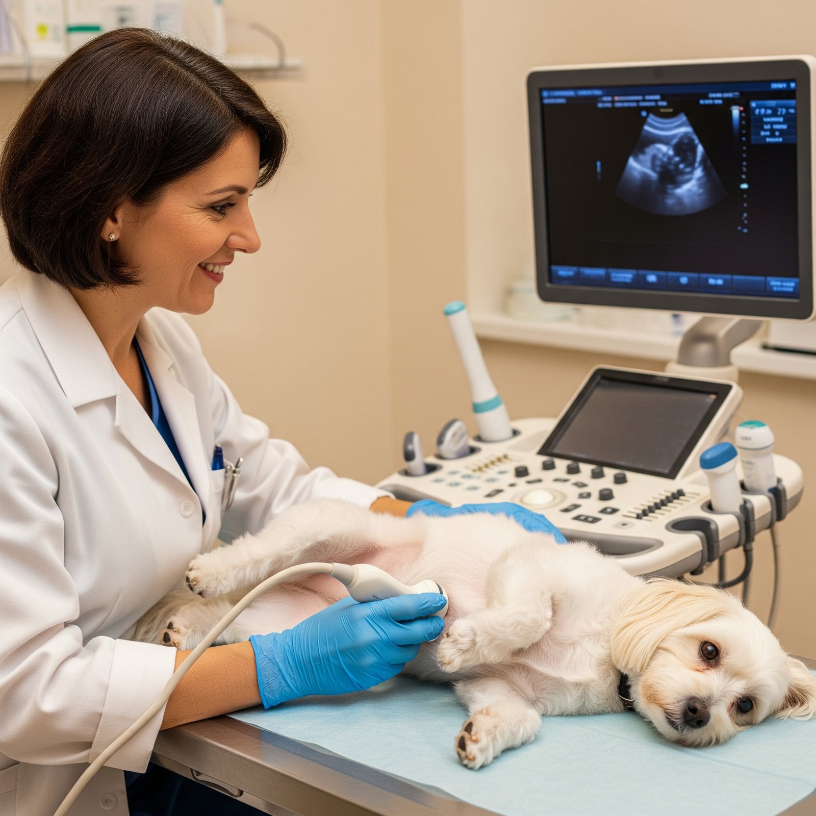
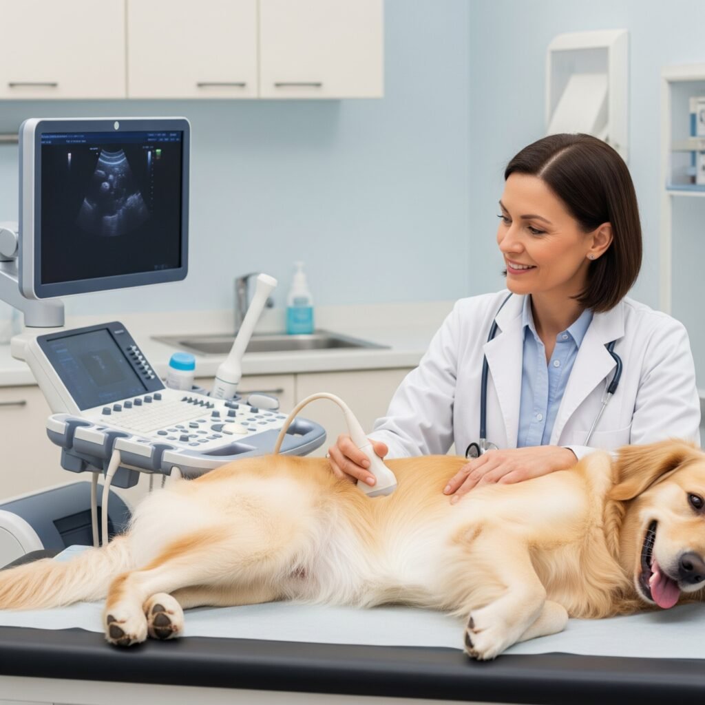
Veterinary Abdominal Ultrasound is one of the most widely used imaging tools in small animal practice, particularly for dogs and cats. Its ability to provide real-time, detailed information about the anatomy and physiology of abdominal organs makes it an indispensable tool for safe, accurate, and minimally invasive diagnostics.
Recent data published in Frontiers in Veterinary Science (2024) indicate that over 80% of general practice veterinarians perform abdominal ultrasounds weekly, both in routine and emergency settings. This underscores the consolidation of ultrasound as a decisive support technology in clinical decision-making.
Clinical Applications of Veterinary Abdominal Ultrasound
Ultrasound is indicated for a wide range of clinical conditions. Its applications include:
- Assessment of abdominal masses, lymph nodes, liver, kidneys, spleen, and bladder
- Diagnosis of gastrointestinal disorders such as intussusceptions or foreign bodies
- Investigation of endocrine diseases like Cushing’s syndrome
- Monitoring of chronic inflammatory conditions (e.g., hepatopathies, enteritis)
- Detection of abdominal effusion or ascites
- Image-guided procedures such as biopsies and fluid aspiration
Studies report that the sensitivity of this exam for detecting structural abdominal changes may exceed 90%, especially when performed by trained professionals using appropriate equipment (PubMed ID: 34510634).
Comparison with Other Diagnostic Tools
Veterinary Abdominal Ultrasound stands out due to its:
- Non-invasive nature, typically not requiring sedation
- Rapid execution, with immediate results
- Safety, with no ionizing radiation exposure
- Repeatability, useful for disease monitoring
Compared to abdominal radiography, ultrasound provides superior evaluation of organ texture, echogenicity, and anatomical boundaries, along with real-time assessment of gastrointestinal motility and urinary flow.
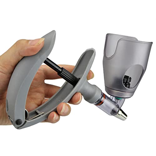
- Continuous Syringe: Automatic spring back design of handle, it is convenient and labor‑saving to use while working.
- Accurate Dose Adjustment: High precise scale dose, adjustable dose, can easy measure the dose you want.
- High quality raw materials: can withstand high temperature and chemical agents, durable and not suitable for breaking.
Role in Pregnancy and Pseudopregnancy
Ultrasound is particularly valuable in reproductive contexts—both for confirming pregnancies and differentiating between clinical and hormonal conditions.
In cases of canine pseudopregnancy—as addressed in the Auquemia Veterinary Guide on False Pregnancy in Dogs—Veterinary Abdominal Ultrasound is instrumental in:
- Confirming the absence of fetal structures
- Detecting uterine fluid or thickening
- Ruling out open or closed pyometra
- Monitoring the progression or resolution of the pseudopregnancy
Its diagnostic accuracy helps clinicians distinguish between hormonal behaviors and pathological conditions, avoiding unnecessary treatments or surgical interventions.

Training and Professional Qualification
Accurate interpretation of Veterinary Abdominal Ultrasound requires solid technical knowledge of anatomy, physiological variations, and abdominal pathologies.
According to Veterinary Radiology & Ultrasound, veterinarians who receive structured POCUS (Point-of-Care Ultrasound) training significantly improve their ability to identify anatomical structures—often doubling their diagnostic accuracy within 12 weeks of supervised practice.
Standardizing scanning protocols and mastering abdominal sweep techniques are key factors in delivering reliable results.
Clinical Benefits for Vets, Pets, and Pet Owners
Routine use of abdominal ultrasound in veterinary clinics provides measurable advantages:
- Faster diagnostic turnaround
- Increased clinical precision
- Reduced need for additional tests
- Higher pet owner confidence and satisfaction
- Optimized treatment plans guided by imaging
Additionally, it enhances the veterinarian’s ability to communicate with pet owners, who can view the exam in real time and better understand their pet’s health status.
Final Considerations
Veterinary Abdominal Ultrasound is now a cornerstone skill in small animal medicine, offering faster, safer, and more accurate diagnosis. Supported by robust scientific literature and applied through standardized protocols, ultrasound contributes decisively to the evolution of modern veterinary practice.
Incorporating this technique into daily clinical routines is not just a differentiator—it’s a professional necessity in the face of growing demand for evidence-based and high-quality animal healthcare.
📚 Main Scientific Sources and Data
- Survey on ultrasound use by small animal veterinarians:
Around 84% have access and 86% use the device multiple times per week
https://pubmed.ncbi.nlm.nih.gov/38668650/ - POCUS hands-on training study (abdominal, thoracic, cardiac):
Improvement from identifying 5.9 to 9.0 anatomical structures in 3 months
https://www.frontiersin.org/articles/10.3389/fvets.2025.1520004 - Regional survey of POCUS use in general veterinary practice (USA):
53% have equipment; 57% use it 4–5+ times per week
https://journals.lww.com/ehpf/fulltext/2018/01020/a_survey_of_point_of_care_ultrasound_use_in.5.aspx - POCUS use in emergency veterinary care:
88% use AFAST and 69% use TFAST protocols
https://www.ncbi.nlm.nih.gov/pmc/articles/PMC7659883/ - Detection of intestinal obstruction with ultrasound vs. radiography:
Ultrasound detected 11/11 cases (100%) vs. 70.7% by X-ray
https://www.veterinary-practice.com/article/examining-the-use-of-diagnostic-imaging-to-identify-intestinal-obstructions